Understanding Electrocardiograms and Stress Tests
What is an electrocardiogram (ECG)?
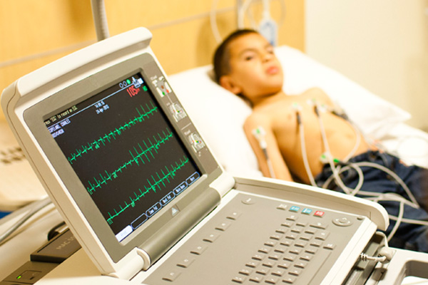
An electrocardiogram (ECG or EKG) is one of the simplest and fastest procedures used to assess the heart. Electrodes (small, plastic patches) are placed at certain locations on the child’s chest, arms and legs. When the electrodes are connected to the ECG machine by lead wires, the electrical activity of the child’s heart is measured. The cardiologist uses this information to decide whether the child needs further tests or the proper course of treatment for the child’s heart problems.
Why is an ECG performed?
The electrical activity of the heart is measured by an electrocardiogram. By placing electrodes at specific locations on the body (chest, arms, and legs), our specialists can get a “picture,” or tracing, of the electrical activity in the heart. Changes in an ECG from the normal tracing can show one, or more, of several heart-related conditions.
Learn more about the electrical system of the heart.
Some medical conditions that can cause changes in the ECG pattern include, but are not limited to, the following:
- Conditions in which the heart is enlarged. These conditions can be caused by various factors, such as congenital (present at birth) heart defects, valve disorders, high blood pressure or congestive heart failure.
- Ischemia. A decreased blood flow to the heart muscle due to clogged or partially-clogged arteries.
- Conduction disorders. A dysfunction in the heart’s electrical conduction system that can make the heart beat too fast, too slow or at an uneven rate.
- Electrolyte disturbances. An imbalance in the level of electrolytes, or chemicals, in the blood, such as potassium, magnesium or calcium.
- Pericarditis. An inflammation or infection of the sack that surrounds the heart.
- Valve disease. Malfunction of one or more of the heart valves that may interfere with blood flow within the heart.
- Chest trauma. Blunt trauma to the chest, such as that suffered in a car accident.
An ECG may also be performed for other reasons, including, but not limited to, the following:
- During a physical examination to obtain a baseline tracing of the heart’s function. (This baseline tracing may be used later as a comparison with future ECGs, to see if any changes have occurred.)
- As part of a workup prior to a procedure, such as surgery, to make sure no heart condition exists that might cause complications during or after the procedure.
- To check the function of an implanted pacemaker.
- To check the effectiveness of certain heart medications.
- To check the heart’s status after a heart-related procedure, such as a cardiac catheterization, heart surgery or electrophysiological studies.
What is the procedure for an ECG?
An ECG can be performed almost anywhere, as the equipment is very compact and portable. At CHOC we provide ECGs in our Heart Center, as well as the inpatient unit of the hospital and the Julia and George Argyros Emergency Department at CHOC. Our specialists even take ECG equipment to
local schools to test athletes. The equipment used includes the ECG machine, skin electrodes and lead wires that attach the electrodes to the ECG machine.
An ECG normally takes approximately five to 10 minutes, including attaching and detaching electrodes. Getting an ECG typically includes the following steps:
- The child lies flat on a table or bed for the procedure.
- The ECG technician uncovers child’s chest only exposing the necessary skin.
- Electrodes (small, plastic patches) are attached to the child’s chest and one electrode is attached to each arm and leg.
- The lead wires are attached to the skin electrodes.
- Once the leads are attached, the technician may key in identifying information such as the child’s name and age into the machine’s computer.
- The ECG is started. The child must lie still and not talk during the procedure, so as not to interfere with the tracing. Caregivers can usually be present in the room and involved in reassuring and encouraging their child during the procedure. At this point, it will take only a few minutes (or less) for the tracing to be completed.
- Once the tracing is completed, the technician will disconnect the leads and remove the skin electrodes.
Depending on the results of the ECG, additional tests or procedures may be scheduled to gather further diagnostic information.
How is the exercise ECG test performed?
At CHOC, stress tests are performed on the third floor of the hospital in our Heart Center. The equipment used includes an ECG machine, electrodes (small, plastic patches that stick on the skin), and lead wires which attach to the skin electrodes. A blood pressure cuff is also used to monitor your child’s blood pressure response during exercise. A treadmill or stationary bicycle is used for exercise.
Each child has an initial, or “baseline,” ECG and blood pressure readings done prior to exercising. He or she will walk on the treadmill or pedal the bicycle during the exercise portion of the procedure. The incline of the treadmill will be gradually increased, or the resistance of the bicycle will be gradually increased, in order to increase the intensity level of exercise. The ECG and blood pressure will be monitored during the exercise portion of the test. Symptoms are also carefully monitored during exercise. The child will be asked to exercise only to the best of his or her ability. Following exercise, ECG and blood pressure readings are monitored for a short time, perhaps another 10 to 15 minutes or so.
The procedure will take approximately one hour, including check-in, preparation and the actual procedure. After the procedure, a hospital stay is not necessary, unless your child’s doctor determines that the child’s condition requires further observation or hospital admission.
The child may feel a little tired or sore for a few hours after the procedure, particularly if he or she is not used to exercising. Otherwise, the child should feel normal within a few hours after the procedure, if not sooner.
Depending on the results of the exercise ECG, additional tests or procedures may be scheduled to gather further diagnostic information.