
Tetralogy of Fallot
Call today to schedule an appointment with one of our pediatric specialists.
What is tetralogy of Fallot and what causes it?
Tetralogy of Fallot (TOF or “TET”) is a heart condition made up of four related congenital (present at birth) defects that is caused due to abnormal development of the fetal heart during the first eight weeks of pregnancy.
The four problems caused by tetralogy of Fallot include:
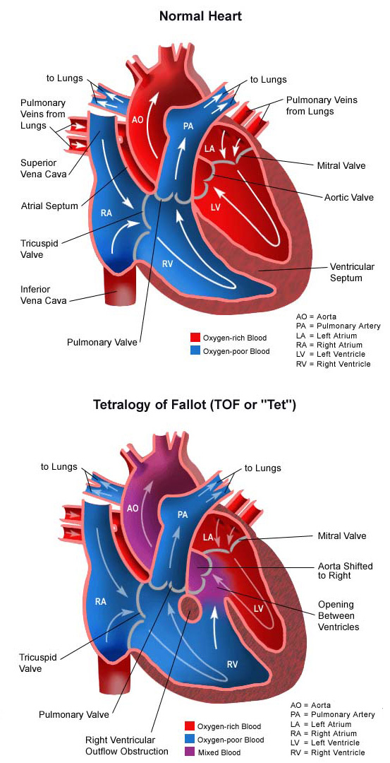
- Ventricular septal defect (VSD). An opening in the ventricular septum, or dividing wall between the two lower pumping chambers of the heart known as the right and left ventricles. Learn more about VSD.
- Pulmonary (or right ventricular outflow tract) obstruction. A muscular obstruction in the right ventricle, just below the pulmonary valve, that decreases the normal flow of blood to the lungs. The pulmonary valve may also be small.
- Overriding aorta. The aorta (main artery leaving the heart) is shifted towards the right side of the heart so that it sits over the ventricular septal defect.
- Right ventricular hypertrophy. The right ventricle becomes thickened as it tries to pump blood past the obstruction into the pulmonary artery.
How does tetralogy of Fallot affect the heart?
Normally, oxygen-poor (blue) blood returns to the right atrium (collecting chamber) from the body, travels to the right ventricle (pumping chamber), then is pumped through the pulmonary artery into the lungs where it receives oxygen. Oxygen-rich (red) blood returns to the left atrium from the lungs, passes into the left ventricle, and then is pumped through the aorta out to the body.
In tetralogy of Fallot, the direction of blood flow within the heart varies, and is largely dependent on the size of the ventricular septal defect, and how severe the obstruction in the right ventricle is.
- With mild right ventricle obstruction, very little of the oxygen-poor (blue) blood in the right ventricle will pass through the VSD to the left ventricle, mix with the oxygen-rich (red) blood there, and then flow into the aorta. The majority of the oxygen-poor (blue) blood will go by its normal route to the lungs. These children may have oxygen levels that are only slightly lower than usual, and do not appear blue.
- With more serious obstruction in the right ventricle, it is harder for oxygen-poor (blue) blood to flow into the pulmonary artery, so more of it passes through the VSD into the left ventricle, mixing with oxygen-rich (red) blood, and then moving on out to the body. These children will have lower than normal oxygen levels in the bloodstream, and may appear blue, especially whenever the pressure in the right ventricle is very high and large amounts of oxygen-poor (blue) blood passes through the VSD to the left side of the heart.
How common is TOF in babies? According to the National Heart, Lung, and Blood Institute, tetralogy of Fallot affects about five of every 10,000 babies and occurs equally in boys and in girls. It is one of the most common congenital abnormalities of the heart that requires intervention in the first year of life.
What are the causes of tetralogy of Fallot?
Some congenital heart defects may have a genetic link causing heart problems to occur more often in certain families. The main causes of TOF are:
Maternal abuse of alcohol during pregnancy, leading to fetal alcohol syndrome, is linked to tetralogy of Fallot. Mothers who take medications to control seizures and mothers with phenylketonuria are also more likely to have a baby with tetralogy of Fallot.
Tetralogy of Fallot may also occur as part of a genetic syndrome like Down syndrome or DiGeorge syndrome.
Most of the time, this heart defect occurs by chance, with no clear reason for its development.
Why is tetralogy of Fallot a concern, and how common is it?
The amount of oxygen-poor (blue) blood that passes through the VSD to the left side of the heart varies. If the right ventricle obstruction is severe, or if the pressure in the lungs is high, a large amount of oxygen-poor (blue) blood passes through the VSD, mixes with the oxygen-rich (red) blood in the left ventricle and is pumped to the body. The more blood that goes through the VSD, the less blood that goes through the pulmonary artery to the lungs, and the less oxygen-rich (red) blood that returns to the right side of the heart.
Some situations, such as crying, increase the pressure in the lungs temporarily, and increasing blueness might be noted as a baby with tetralogy of Fallot cries. In other situations, the pathway from the right ventricle to the pulmonary artery becomes tighter, preventing much blood from passing that way, and allowing oxygen-poor (blue) blood to flow through the VSD into the left heart circulation. Both of these situations are nicknamed “TET spells.” Usually, TET spells resolve after the baby calms down, but on rare occasions, they may continue or get worse, which can be a medical emergency. Sometimes, steps can be taken to lessen the pressure or the obstruction, and allow more blood to flow into the lungs and less through the VSD. These steps, however, are not always effective.
What are the symptoms of tetralogy of Fallot?
The following are the most common symptoms of tetralogy of Fallot. However, each child may experience symptoms differently.
- Because large amounts of oxygen-poor (blue) blood can flow to the body under certain circumstances, one of the symptoms of tetralogy of Fallot is blueness (blue color of the skin, lips and nail beds) that occurs with such activity as crying or feeding.
- Some babies do not have noticeable cyanosis (blue color of the skin, lips and nailbeds), but may instead be very irritable or lethargic due to a reduced amount of oxygen in the bloodstream.
- Some children become pale or ashen in color, and may have cool, clammy skin.
Any of these can be symptoms of tetralogy of Fallot. The TOF symptoms may resemble other medical conditions or heart problems. If you identify any of these symptoms, consult the child’s doctor for a proper diagnosis.
How is tetralogy of Fallot in children diagnosed?
If a child’s doctor detects a murmur during a physical examination, the child may be referred to a pediatric cardiologist for a diagnosis. If the heart murmur is caused by the turbulence of blood flowing through the obstruction from the right ventricle to the pulmonary artery, the cardiologist will diagnose the child with tetralogy of Fallot. The child’s symptoms also help with the diagnosis.
A pediatric cardiologist specializes in the diagnosis and medical management of congenital heart defects, as well as heart problems that may develop later in childhood. The cardiologist will perform a physical examination, listen to the heart and lungs, and make other observations that help in the diagnosis. The location within the chest that the murmur is heard best, as well as the loudness and quality of the murmur (such as, harsh or blowing) will give the cardiologist an initial idea of which heart problem the child may have. Diagnostic testing for congenital heart disease varies by the child’s age, clinical condition and institutional preferences. Some tests that may be recommended include the following:
What is the treatment for tetralogy of Fallot?
Specific treatment for tetralogy of Fallot is determined by the child’s healthcare team based on:
- The child’s age, overall health and medical history
- Extent of the condition
- The child’s tolerance for specific medications, procedures or therapies
- Expectations for the course of the condition
- The family’s opinion or preference.
Tetralogy of Fallot is treated by surgical repair of the defects. A team of cardiac surgeons does the surgery, usually around 6 months of age, or even earlier, if the baby is having problems with TET spells. Repairing the heart defects will allow oxygen-poor (blue) blood to travel its normal route through the pulmonary artery to receive oxygen. The TOF surgery is performed under general anesthesia and involves closing the ventricular septal defect with a patch and opening up the blocked pathway between the right ventricle and the pulmonary artery is with a patch. If the pulmonary valve is small, it may be opened as well.
Tetralogy of Fallot surgery and recovery: What can families expect?
Children who undergo treatment for tetralogy of Fallot spend time in the cardiovascular intensive care unit (CVICU) after the repair. During the first several hours after surgery, the child will be very drowsy from the anesthesia that was used during the operation, and from medications are given to relax him or her and to help with pain. As time goes by, the child becomes more alert.
While the child is in the CVICU, special equipment is used to aid in recovery and may include the following:
- Ventilator. A machine that helps the child breathe while he or she is under anesthesia during the operation. A small, plastic tube is guided into the windpipe and attached to the ventilator, which breathes for the child while he or she is too sleepy to breathe on his or her own. After a tetralogy of Fallot repair, some children may remain on the ventilator for up to several days so they can rest, while others may be able to breathe on their own within a few hours of the surgery. The hospital care team will provide families with information on how long they expect the child to need a breathing tube.
- Intravenous (IV) catheters. Small, plastic tubes inserted through the skin into blood vessels to provide IV fluids and important medicines that help the child recover from the operation.
- Arterial line. A specialized IV placed in the wrist or other area of the body where a pulse can be felt, that measures blood pressure continuously during surgery and while the child is in the CVICU.
- Nasogastric (NG) tube. A small, flexible tube that keeps the stomach drained of acid and gas bubbles that may build up during surgery.
- Urinary catheter. A small, flexible tube that allows urine to drain out of the bladder and accurately measures how much urine the body makes, which helps determine how well the heart is functioning. After surgery, the heart may be a little weaker than it was before, and the body may start to hold onto fluid, causing swelling and puffiness. Diuretics (medicine to increase urine volume) may be given to help the kidneys remove excess fluid from the body.
- Chest tube. A drainage tube will be inserted inside the chest to drain blood and fluid that would otherwise accumulate after the incision is closed. Bleeding may occur for several hours, or even a few days after surgery.
- Heart monitor. A machine that constantly displays a picture of the child’s heart rhythm, and monitors heart rate, arterial blood pressure and other values.
Some patients may need other equipment not mentioned while in the CVICU or afterwards. Hospital staff will explain all of the equipment necessary for recovery as needed.
Patients are kept as comfortable as possible with several different medications—some relieve pain and relieve anxiety. Hospital staff will ask the child’s parents for their input on how to best comfort and soothe the child.
As the child recovers, they will begin eating, being out of the bed in a parent’s lap or walking (if they are old enough). Before the child is discharged from the hospital, parents will learn how to care for the child at home. The staff will provide instructions regarding medications, activity limitations and follow-up appointments before the child is discharged.
How is a child cared for at home after tetralogy of Fallot surgery?
Tips for TOF surgery recovery: Pain medications, such as acetaminophen or ibuprofen, may be recommended to keep the child comfortable at home. The child’s doctor will discuss pain control before the child is discharged from the hospital.
After TOF surgery, older children can usually handle light activity for a short amount of time. Children usually become tired easily and sleep more than before surgery. Within a few weeks, however, the child should be fully recovered.
What is the long-term outlook for children diagnosed with tetralogy of Fallot?
Most children who have had a tetralogy of Fallot surgical repair live healthy lives. Activity levels, appetite and growth return to normal in most children soon after surgery. The child’s cardiologist may recommend that antibiotics be given to prevent bacterial endocarditis after discharge from the hospital.
After initial repair of tetralogy of Fallot, pulmonary valve replacement may be needed as the child nears or reaches adulthood to prevent complications, such as enlargement of the right ventricle, abnormal heart rhythms and heart failure. In adulthood, women considering having a baby should speak with their cardiologist before getting pregnant.
The child’s healthcare team will provide more details on the specific long-term outlook for the children.