
Aortic Stenosis
What is aortic stenosis?
Aortic stenosis is a heart defect that may be congenital (present at birth) or acquired (develop later in life). If the problem is congenital, then something started to occur during the first 8 weeks of pregnancy to affect the development of the aortic valve. Congenital aortic stenosis occurs three times more often in boys than in girls.
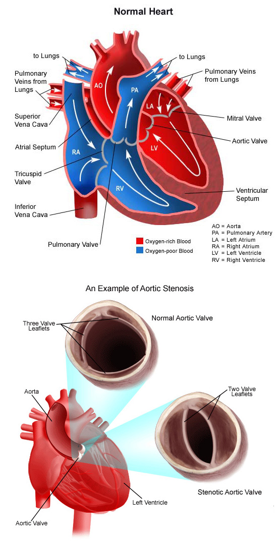
The aortic valve is found between the left ventricle and the aorta. It has three leaflets that function like a one-way door, allowing blood to flow forward into the aorta, but not backward into the left ventricle. Aortic stenosis is the inability of the aortic valve to open completely.
With aortic stenosis, problems with the aortic valve make it harder for the leaflets to open and permit blood to flow forward from the left ventricle to the aorta. In children, these problems can include a valve that:
- Only has two leaflets instead of three (bicuspid aortic valve).
- Has leaflets that are partially fused together.
- Has thick leaflets that do not open all the way.
- Becomes damaged by rheumatic fever or bacterial endocarditis.
- Area above or below the valve is narrowed (supravalvar or subvalvar).
Aortic stenosis may be present in varying degrees, graded according to how much obstruction to blood flow is present. A child with severe aortic stenosis will be quite ill, with major symptoms noted early in life. A child with mild aortic stenosis may have few symptoms, or perhaps none until later in adulthood. The degree of obstruction can become worse with time.
What causes aortic stenosis?
Congenital aortic stenosis occurs due to improper development of the aortic valve in the first 8 weeks of fetal growth. It can be caused by a number of factors, though most of the time, this heart defect occurs sporadically (by chance), with no apparent reason for its development.
Some congenital heart defects may have a genetic link, either occurring due to a defect in a gene, a chromosome abnormality, or environmental exposure, causing heart problems to occur more often in certain families.
Acquired aortic stenosis may occur after a strep infection that progresses to rheumatic fever.
Why is aortic stenosis a concern?
Mild aortic stenosis may not cause any symptoms. Moderate to severe aortic stenosis, however, can present in a number of several different ways, including:
- The left ventricle has to work harder to try to move blood through the tight aortic valve. Eventually, the left ventricle is no longer able to handle the extra workload, and it fails to pump blood to the body efficiently.
- There is a higher than average chance that the aorta may become dilated (enlarged). This can increase the risk of an aneurysm or dissection of the aorta.
- There is a higher than average chance of developing an infection in the lining of the heart or aorta known as bacterial endocarditis.
- The coronary arteries, which supply oxygen-rich (red) blood to the heart muscle, may not receive enough blood to meet the demands of the heart.
What are the symptoms of aortic stenosis?
The following are the most common symptoms of aortic stenosis. However, each child may experience symptoms differently. Symptoms may include:
- Fatigue
- Dizziness with exertion
- Shortness of breath
- Irregular heartbeats or palpitations
- Chest pain.
The symptoms of aortic stenosis may resemble other medical conditions or heart problems. Always consult the child’s physician for a diagnosis.
How is aortic stenosis diagnosed?
If a child’s physician hears a heart murmur during a physical examination, he or she will refer the child to a pediatric cardiologist for a diagnosis. A heart murmur is a noise caused by the turbulence of blood flowing through the obstruction from the left ventricle to the aorta. Symptoms your child exhibits will also help with the diagnosis.
CHOC pediatric cardiologists specialize in the diagnosis and medical management of congenital heart defects and heart problems that may develop later in childhood. To diagnose aortic stenosis, the cardiologist will perform a physical examination by listening to the child’s heart and lungs and make other observations that help in the diagnosis. The loudness and quality of the murmur (harsh, blowing, etc.) as well as the location within the chest that the murmur is heard best will give the cardiologist an initial idea of which heart problem your child may have. Diagnostic testing for congenital heart disease is determined by the child’s age and clinical condition. Some tests that may be recommended include the following:
- Chest X-ray. A diagnostic test that uses X-ray beams to produce images of internal tissues, bones and organs onto film. Learn more about chest X-rays.
- Electrocardiogram (ECG or EKG). A test that records the electrical activity of the heart, shows abnormal rhythms (arrhythmias or dysrhythmias), and detects heart muscle stress. Learn more about electrocardiogram.
- Echocardiogram (echo). A procedure that evaluates the structure and function of the heart by using sound waves recorded on an electronic sensor that produce a moving picture of the heart and heart valves. Learn more about echocardiogram.
- Exercise electrocardiogram (ECG or EKG). An exercise EKG is done to assess the heart’s response to stress or exercise. The EKG is monitored while your child is exercising on a treadmill or stationary bike. An EKG measures the electrical activity of your child’s heart. Learn more about exercise EKG.
- Cardiac MRI. A diagnostic procedure that uses a combination of large magnets, radiofrequencies and a computer to produce detailed images of the heart. Learn more about MRI.
How is aortic stenosis treated?
Specific treatment for aortic stenosis is determined by the child’s physician based on:
- The child’s age, overall health and medical history
- Extent of the aortic stenosis
- The child’s tolerance for specific medications, procedures or therapies
- Expectations for the course of the aortic stenosis
- The family’s opinion or preference.
Aortic stenosis is treated with repair of the obstructed valve. Several options are currently available.
Some infants who are very sick require care in the cardiovascular intensive care unit or the neonatal intensive care unit prior to the repair procedure. Others who are exhibiting few symptoms will have the repair scheduled on a less urgent basis.
Activity may be limited in children who have moderate aortic stenosis prior to repair. For instance, competitive sports that require endurance may be restricted.
Your child’s cardiologist and heart surgeon will carefully review your child’s case and recommend one of several treatment options, based on what treatment they think is the best for your child’s heart problem. Repair options include the following:
- Balloon dilation. A cardiac catheterization procedure in which a small, flexible tube (catheter) is inserted into a blood vessel in the groin, and guided to the inside of the heart. The tube has a deflated balloon in the tip. When the tube is placed in the narrowed valve, the balloon is inflated to stretch the area open.
- Valvotomy. A heart surgery procedure in which a surgical release is done to calcified tissue within the aortic valve leaflets that are preventing the valve leaflets from opening properly.
- Aortic valve replacement. The aortic valve is replaced with a new valve with a surgical procedure. Replacement valves fall into two categories: Tissue (biological) valves, which are made from animal tissues and valves that are soft and thin. Mechanical valves, which are made from carbon fibers, have solid valve leaflets. Children who have undergone a valve replacement will need to follow antibiotic prophylaxis throughout their lifetime. Patients who have received a mechanical valve will need life-long treatment with Coumadin (warfarin), a medication that thins the blood to prevent catastrophic clots from forming on the valve leaflets themselves.
- Aortic homograft. A section of aorta from a human donor with its aortic valve intact is used to replace the aortic valve and a section of the ascending aorta. This procedure is seldom performed due to the complexity of the operation and the rapid failure of the aortic homograft itself.
- Pulmonary autograft (Ross procedure). A section of the child’s own pulmonary artery with the pulmonary valve intact is used to replace the aortic valve and a section of the ascending aorta. A section of pulmonary artery from another human donor with its valve intact is used to replace the transferred pulmonary artery.
What is the long-term outlook after aortic stenosis surgical repair?
Most children who have had an aortic stenosis surgical repair will live healthy lives. Activity levels, appetite and growth should eventually return to normal.
As the child grows, a valve that was ballooned may once again become narrowed. If this happens, a second balloon procedure or operation may be necessary to repair aortic stenosis. Sometimes the aortic tissue itself may be abnormal, which might lead to complications in the teen or adult years. Regular follow-up care at a specialized cardiac center should continue throughout life.
Individuals who had a mechanical valve replacement may need to take anticoagulants (blood thinners) to prevent blood clots from forming on the artificial valve surfaces. It is extremely important that your child receives this medication on a regular basis and gets regular monitoring of the blood’s clotting status in order to maintain the most appropriate dose of anticoagulants. Because children grow so rapidly, medication doses change often. It is important to keep regular appointments with your child’s cardiologist to make sure your child receives safe and effective medication doses.
Initial valve replacement is often performed using a tissue valve to avoid the need for anticoagulation, especially for females of childbearing age. Anticoagulation during pregnancy is very difficult to manage, and requires special treatment.
Repeat valve replacement is not uncommon during a person’s lifespan, especially when replacement happens in a growing child. In addition, blood pressure should be closely monitored and managed.
The cardiologists at the Heart Institute will discuss your child’s specific long-term outlook.
What to Expect After Aortic Stenosis Surgical Repair
Children who require surgery will go to the cardiovascular intensive care unit (CVICU) after the procedure. While a child is in the CVICU, special equipment will be used to help him or her recover from surgery, and may include the following:
- Ventilator: A machine that helps your child breathe while he or she is under anesthesia during the operation. A small, plastic tube is guided into the windpipe and attached to the ventilator, which breathes for your child while he or she is too sleepy to breathe effectively on his or her own. Many children remain on the ventilator for a few hours to a few days after surgery so they can rest.
- Intravenous (IV) catheters. Small, plastic tubes inserted through the skin into blood vessels to provide IV fluids and important medicines that help your child recover from the operation.
- Arterial line. A specialized IV placed in the wrist, or other area of the body where a pulse can be felt, that measures blood pressure continuously during surgery and while your child is in the CVICU.
- Nasogastric (NG) tube. A small, flexible tube that keeps the stomach drained of acid and gas bubbles that may build up during surgery.
- Urinary catheter. A small, flexible tube that allows urine to drain out of the bladder and accurately measures how much urine the body makes. This can help determine how well the heart is functioning. After surgery, the heart may be a little weaker than it was before, and, therefore, the body may start to hold onto fluid, causing swelling and puffiness. Diuretics may be given to help the kidneys remove excess fluid from the body.
- Chest tube. A drainage tube may be inserted to keep the chest free of blood that would otherwise accumulate after the incision is closed. Bleeding may occur for several hours, or even several days after surgery.
- Heart monitor. A machine that constantly displays a picture of your child’s heart rhythm, and monitors heart rate, arterial blood pressure, and other values.
Other equipment, not mentioned here, may be used in the CVICU and will explained as needed.
Following a procedure or surgery, children are kept as comfortable as possible with several different medications; some of which relieve pain and some of which relieve anxiety. Our staff, including our certified child life specialists, work with the family to find the best ways to soothe and comfort the child.
As your child recovers, you will learn how to care for your child at home before your child is discharged. Your child may need to take medications for a while, and these will be explained to you. The staff will give you instructions regarding medications, activity limitations and follow-up appointments before your child is discharged. Your child’s cardiologist may recommend that antibiotics be given to prevent bacterial endocarditis after discharge from the hospital.