
Echocardiogram
Call today to schedule an appointment with one of our pediatric specialists.
Cardiology & Cardiothoracic Surgery Referrals
Physicians can refer patients to CHOC through our eCeptionist Referral Portal.
View the referral guidelines
Dr. Wyman Lai, pediatric cardiologist, discusses why a child might need an echocardiogram of the heart, what it is and how it is done. Learn more about the CHOC Heart Institute.
Understanding Echocardiograms
If a doctor thinks a patient may have a heart defect, heart disease or other heart problem, he or she may suggest the child have a test called an “echocardiogram.” Echocardiograms are common, safe procedures that help doctors look at how the heart is working. They may also be called an “echo,” “cardiac ultrasound,” “ultrasonography” or “cardiac Doppler.”
Echocardiography is another word that families may hear used by health care providers. Echocardiography refers to the overall science or discipline of looking at the heart’s structures and functions while using different types of electrocardiogram tests. At CHOC we have a team of expert echocardiographers experienced in working with children of all ages and levels of development.
There are two types of echocardiograms: traditional, transthoracic echocardiograms and transesophageal echocardiograms.
Frequently Asked Questions
Because parents often have a lot of questions about echocardiograms, the experts at the CHOC Heart Institute have put together answers to some of the most frequently asked questions patients and their families ask about echocardiography.
What is an echocardiogram?
An echocardiogram is a non-invasive procedure used to assess the heart’s structure and function (a heart ultrasound). During the procedure, a transducer (like a microphone) sends out ultrasonic sound waves at a frequency too high to be heard. When the transducer is placed at certain locations and angles, the ultrasonic sound waves move through the skin and other body tissues to the heart tissues, where the waves bounce or “echo” off of the heart structures. The transducer picks up the reflected waves and sends them to a computer. The computer displays the echoes as images of the heart walls and valves. Transthoracic echocardiograms (TTE) are the most common type of echocardiograms performed.
An echocardiogram may utilize several special types of echocardiography, as listed below:
- M-mode echocardiography. This, the simplest type of echocardiography, produces an image that is similar to a tracing rather than an actual picture of heart structures. M-mode echo is useful for measuring heart structures, such as the heart’s pumping chambers, the size of the heart itself and the thickness of the heart walls.
- Doppler echocardiography. This Doppler technique is used to measure the flow of blood through the heart’s chambers and valves. The amount of blood pumped out with each beat helps the doctor understand how the heart is working. Doppler can also help doctors find problems with the heart’s valves or walls by looking for abnormal blood flow within the heart.
- Color Doppler. In color Doppler, different colors are used by the computer to show the direction of blood flow. This makes using Doppler echocardiography much easier.
- 2-D (two-dimensional) echocardiography. This technique is used to “see” the actual motion of the heart structures. On a special monitor, the heart appears as a cone shape and the doctor can watch the real-time motion of all of the different parts of the patient’s heart. By seeing how the different parts of the heart are working, the doctor can get a better understanding of the patient’s heart problems.
- 3-D (three-dimensional) echocardiography. 3-D echo technique captures three-dimensional views of the heart structures with even more detail than 2-D echo. The live images allow the doctor to measure the heart’s functions while it is beating, which makes it easier for the doctor to diagnose complex heart problems and create treatment plans for heart disease without the use of cardiac catheterization.
A second type of echocardiogram used is a transesophageal echocardiogram (TEE). TEE uses the same technology as TTE, but TEE is invasive (goes inside the body). Learn more about transesophageal echocardiogram.
Why is an echocardiogram performed?
Echocardiograms are used to help doctors better diagnose a variety of heart conditions, including:
- Cardiomyopathy. An enlargement of the heart due to thickening or weakening of the heart muscle. Learn more about cardiomyopathy.
- Congenital heart defects. Defects in one or more heart structures that occur during formation of the fetus, such as a ventricular septal defect (hole in the wall between the two lower chambers of the heart). Learn more about congenital heart defects.
- Congestive heart failure. A condition in which the heart muscle has become weakened to an extent that blood cannot be pumped efficiently, causing fluid buildup (congestion) in the blood vessels and lungs and edema (swelling) in the feet, ankles and other parts of the body. Learn more about congestive heart failure.
- Aneurysm. A dilation of a part of the heart muscle or the aorta (the large artery that carries oxygenated blood out of the heart to the rest of the body), which may cause weakness of the tissue at the site of the aneurysm.
- Valvular heart disease. Malfunction of one or more of the heart valves that may cause an abnormality of the blood flow within the heart.
- Cardiac tumor. A tumor of the heart that may occur on the outside surface of the heart, within one or more chambers of the heart (intracavitary), or within the muscle tissue (myocardium) of the heart.
- Pericarditis. An inflammation or infection of the sac that surrounds the heart.
An echocardiogram may also be simply performed to assess the heart’s overall function and general structure.
What are the risks of undergoing a transthoracic (standard) echocardiogram?
For some patients undergoing transthoracic echocardiograms (TTE), having to lie still on the examination table for the length of the procedure may cause some discomfort or pain.
There may be other risks depending on the child’s specific medical condition. Be sure to discuss any concerns with the child’s doctor prior to the procedure.
Learn about the risks associated with a more invasive transesophageal echocardiogram (TEE).
How to patients prepare for an echocardiogram?
The child’s doctor will explain the echocardiogram test before the day of the procedure. This is a good time for the child and family to ask the doctor questions.
Generally, no prior preparation, such as fasting or sedation, is required. Patients or parents should, however, be certain to tell the child’s doctor about all medications (prescription and over-the-counter) and herbal supplements the child takes and any other medical conditions the child may have.
What happens during an echocardiogram?
An echocardiogram may be performed on an outpatient basis or as part of a child’s stay at CHOC. The procedure may vary depending upon the child’s condition.
Generally, an echocardiogram follows this process:
- The child is asked to remove any jewelry or other objects that may interfere with the procedure. Patients do not need to remove glasses or hearing aids. The child is also asked to remove clothing from the waist up and given a gown to wear.
- The patient is then asked to lie on an exam table or bed. A pillow or wedge may be placed behind the child’s back for support.
- Once properly positioned, the child is connected to an ECG monitor that records the electrical activity of the heart and monitors the heart during the procedure using small, adhesive electrodes. The ECG tracings that record the electrical activity of the heart will be compared to the images displayed on the echocardiogram monitor. The room is darkened so that the images on the echo monitor can be viewed by the sonographer.
- The sonographer will place warmed gel on the child’s chest and then place the transducer probe on the gel. The child will only feel slight pressure as the sonographer positions the transducer to obtain the desired images of the heart.
- During the test, the sonographer will move the transducer probe around and apply varying amounts of pressure to obtain images of different locations and structures of your heart. The amount of pressure used with the probe to get images should not be uncomfortable. The sonographer will ensure the patient is comfortable during test, but patients are encouraged to inform the sonographer if pain or discomfort are experienced during the procedure.
- After the procedure has been completed, the sonographer will wipe the gel from the child’s chest and remove the ECG electrode pads. The child may redress.
In the case of a stress or exercise echocardiogram, the child will have the resting test before and after exercise on a treadmill or stationary bike.
What is to be expected after an echocardiogram?
There are usually no special instructions after an echocardiogram and patients can resume their normal diet and activities soon after the procedure. In some cases, however, the doctor may provide additional or alternate instructions after the procedure, depending on the child’s particular situation and medical condition.
Understanding Transesophageal Echocardiogram
What is a transesophageal echocardiogram?
A transesophageal echocardiogram (TEE) is a diagnostic procedure used to assess the heart’s structure and function (a heart ultrasound). Just like a traditional transthoracic echocardiogram, a transducer sends out ultrasonic sound waves at a frequency too high to be heard. When the transducer is placed at certain locations and angles, the ultrasonic sound waves move through the skin and other body tissues to the heart tissues, where the waves bounce or “echo” off of the heart structures. The transducer picks up the reflected waves and sends them to a computer. The computer displays the echoes as images of the heart walls and valves.
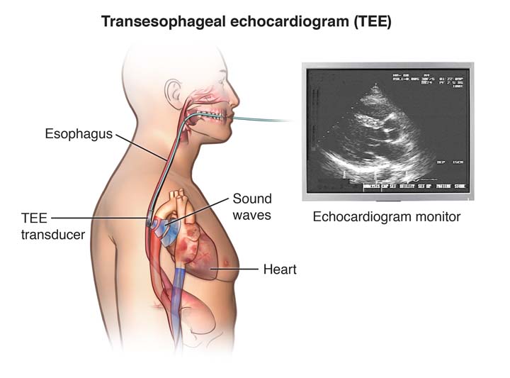
A TEE is performed by inserting a probe with a transducer down the esophagus, rather than placing the transducer on the chest as in a standard transthoracic echocardiogram (TTE). Because the probe is in the esophagus which is located right behind the heart, TEE provides a clearer images.
Certain conditions of the heart, such as valve disorders, implanted prosthetic (artificial) heart valves, and blood clots or masses inside the heart, are often better seen with TEE.
TEE is usually used during heart surgery to further assess the heart structure and function both before and after the operation to help optimize the heart surgery and make it safer.
Like a TTE, TEE may utilize one or more of several special types of echocardiography, including M-mode, Doppler, color Doppler, as well as 2-D (two-dimensional) and 3-D (three-dimensional) technologies.
What are the risks of the transesophageal echocardiogram?

Possible risks associated with a transesophageal echocardiogram include, but are not limited to, the following:
- Breathing problems
- Heart rhythm problems
- Infection
- Bleeding
- Injury to the esophagus, throat or inside of the mouth.
Patients with known problems of the esophagus, such as esophageal varices, esophageal obstruction or radiation therapy to the area of the esophagus should be evaluated carefully by the doctor before having the procedure.
There may be other risks depending upon a child’s specific medical condition or health history. Be sure to discuss any concerns with the child’s doctor prior to the procedure.
CHOC is accredited by the Intersocietal Accreditation Committee for pediatric transesophageal echocardiogram. IAC accreditation ensures a commitment to the quality of health care provided to patients.
What happens before a transesophageal echocardiogram?
- Before the procedure, the child’s doctor will explain the procedure and provide family members and the patient the opportunity to ask questions.
- The child’s legal guardian will be asked to sign a consent form that gives the doctor permission to do the test. It is important to read the form carefully and ask questions if something is not clear.
- The patient will need to fast (not eat or drink) for a certain period of time prior to the procedure. (This is often referred to as “NPO.”) The child’s doctor will provide specific guidelines. Learn more about NPO at CHOC.
- Parents should notify their child’s doctor if the child is allergic to or sensitive to medications, local anesthesia or latex. It is also important to tell the child’s doctor about all medications (prescription and over-the-counter) and herbal supplements the child may be taking, and if the child has a history of bleeding disorders, takes any anticoagulant (blood-thinning) medications, or other medications that affect blood clotting. The child’s doctor will provide instruction on which medications and supplements can and cannot be taken prior to the procedure.
- Prior to the TEE, the doctor may request a blood test prior to determine how long it takes for the child’s blood to clot. Other blood tests may be done as well.
- Patients are given sedative prior to the procedure to help them relax.
- Based on the child’s medical condition, other preparations may need to made before the procedure.
What happens during a transesophageal echocardiogram?
A TEE may be performed on an outpatient basis or as part of a stay in at CHOC. Procedures may vary depending on the child’s specific condition.
All patients are admitted to the Tidwell Procedure Center at CHOC. After being taken to a preoperative room, the child will:
- Remove jewelry or other objects that may interfere with the procedure.
- Remove clothing and put on a hospital gown.
- Use the restroom.
- Have an intravenous (IV) line placed in their hand or arm prior to the procedure so that medications and fluids can be given to the child as needed. The child is given a sedative through the IV that will help them feel more relaxed.
Once the child has been prepared for surgery, the doctor will answer any last minute questions before the child is taken to the operating room for the TEE. An anesthesiologist will usually assist with this procedure.
Once inside the room, the child lies on a bed. A pillow or wedge may be placed behind the child’s back for support and the child is connected to a cardiorespiratory monitor that records the electrical activity of the heart and monitors the heart during the procedure using small, adhesive electrodes. The child’s vital signs (heart rate, blood pressure, breathing rate and oxygenation level) are also monitored during the procedure. Some patients may also receive oxygen through nasal tubes.
After making sure the child is properly positioned and connected to the monitoring equipment, a local anesthetic spray is applied to the back of the throat. This spray numbs the back of the throat and makes passing the TEE probe more comfortable.
When the procedure begins, the TEE probe is very carefully passed through the child’s mouth and down the throat by the cardiologist. Once the probe is in the right place, the doctor will be able to see images of the structure of the child’s heart. After the necessary images are obtained, the probe is removed from the child’s throat. The procedure usually takes 20-30 minutes, depending on the child’s specific condition.
What happens after a transesophageal echocardiogram procedure?
After the procedure, the child is moved to the recovery area of the Tidwell Procedure Center. It is also referred to as the “PACU.” There, nurses monitor the child’s heart rate, ECG, blood pressure and oxygen levels. Once the child is alert, their gag reflex has returned and vital signs are normal, the ECG electrode pads, the oxygen probe and the IV will be removed. A nurse will let the child’s parents know when they can see them in the recovery area.
Some children may feel weak, tired or groggy for the remainder of the day of the procedure, and should feel normal by the day after the procedure. The patient’s voice maybe horse and/or their throat may be sore for a few days following the procedure due to the insertion of the TEE probe. This is normal but if it persists or worsens the child may need to be evaluated by a doctor.
Usually, children under the age of 1 year will be admitted to the hospital overnight to the cardiovascular intensive care unit (CVICU) for monitoring, while most other children can go home the same day.
The child should be able to resume his or her regular diet and activities the same or next day unless the doctor advises differently.
Generally, there is no special type of care following a TEE. However, the child’s doctor may have additional or alternate instructions after the procedure, depending on the child’s particular situation.